These muscles are primarily responsible for extending. Learning Objectives Outline the posterior muscles of the torso.
![]()
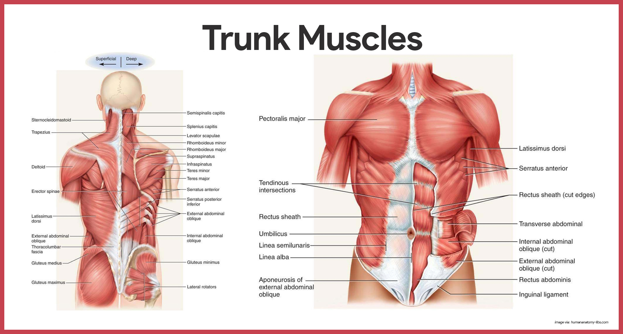
Muscles Of The Trunk Anatomy Diagram Pictures Kenhub
There is a printable worksheet available for download here so you can take the quiz with pen and paper.

. The musculature of your trunk or core is of vital importance to understand fully from both a stability perspective when trying to understand problems like back pain and weakness and also crucial to understand when trying to execute many of your bodys primary movements. The clinical aspect of the anatomy contained in the posterior neck triangle is useful for a wide variety of medical specialties including anesthesiology otolaryngology physical medicine and rehabilitation and others. The diaphragm is another muscle in the thorax that serves as the main muscle of inspiration.
The muscle located closest to the spine. The erector spinae group of muscles on each side of the vertebral column is a large muscle mass that extends from the sacrum to the skull. Muscles of the Trunk.
The legs include the upper leg knee lower leg ankle and. Muscles anatomy muscular system. The suboccipital muscles are located deep to trapezius in the suboccipital region of.
-Form part of the posterior abdominal wall. Google Classroom Microsoft Teams LTI. Terms in this set 8 Deltoid.
This blog post article is an overview of the muscles of the pelvis. Trunk Anatomy Stability. Identify the posterior trunk muscles described in Column A by choosing a response from Column B.
Superficial muscles of the back. View 08-muscles-Anatomy-of-anterior_trunkppt from ALL 192 at University of Nairobi. Figures 8-5 and 8-6 shows many of the muscles of the bodys trunk that you need to know as well as some of the muscles of the arms and legs you will learn about in the next lab.
Similar to learning the muscles of the lumbar spinetrunk it can be. 2017 Elsevier should be consulted. The muscle in blue is the.
Deep muscles of the back. Create your own free activity from our activity creator. For more complete coverage of the structure and function of the low back and pelvis The Muscular System Manual The Skeletal Muscles of the Human Body 4th ed.
The posterior neck triangle is a clinically relevant anatomic region that contains many important vascular and neural structures. Select a different color for each muscle description with a coding circle and color the ccxling circles and corresponding muscles on Figure 68. For descriptive purposes the muscles of the back are divided into two groups.
Most superficial muscle of posterior thorax flat and triangular in shape O. Muscles of the Pelvis. What is at A.
The muscles of the trunk include those that move the vertebral column the muscles that form the thoracic and abdominal walls and those that cover the pelvic outlet. Large rounded triangular shoulder muscle. Your Skills Rank.
Posterior Trunk Muscles Posterior Trunk Muscles Infraspinatus Splenius Capitus Latissimus Dorsi Rhomboid Minor Rhomboid Minor Illiocostalis Gluteus Medius Longissimus Trapezius Levator Scapulae Deltoid Rhomboid Major Internal Abdominal Oblique Internal Abdominal. Click to Rate Hated It Click to Rate Didnt Like It Click to Rate Liked It Click to Rate Really Liked It Click to Rate Loved It 45 1. The back is the body region between the neck and the gluteal regions.
Anatomy anatomy-physiology undergraduate1. It is the most superficial of the calf muscles. Movement patterns are complex and most muscles func-LEVER SYSTEMS lever Anatomy of the Muscular System Chapter 10 D.
The muscles of the anterior thorax provide movements to the arm and shoulder while the muscles of the posterior thorax also help change thoracic volume during breathing and reinforce the thoracic wall. -Acting separately each muscle of the pair flexes the spine laterally. Myology Bony Anatomy of the Thorax 022722 1 Gross Anatomy Osteology of the Thorax 022722 2 Thorax Thorax.
This muscle of the abdomen has fibers that run perpendicular to the midline and works to compress the abdomen. Extends the trunk and laterally bends the trunk rotates the trunk to the opposite side dorsal primary rami of spinal nerves C1-T12 supplied segmentally by. This muscle is the prime mover in arm abduction.
Both heads attach to the back surface of the calcaneus also. S Column A 1. Posterior trunk muscles Suboccipital muscles of the neck.
Muscles posterior Trunk label Map Quiz 5 muscles. The legs are the lower limbs of the human body that provide support and stability in addition to allowing movement. Enter the correct letter in rhe answer blank.
It comprises the vertebral column spine and two compartments of back muscles. -Acting together they extend the lumbar spine. Diamond-shaped muscle of posterior neck and upper back that extends from the skull to spine of scapula to vertebral column.
Deep cervical a posterior intercostal aa subcostal aa lumbar aa. Trunk Muscles 289 Muscles of the Thorax 289 Muscles of the Abdominal Wall 289 Muscles of the Back 290 Muscles of the Pelvic Floor 290 Upper Limb Muscles 293. This is an online quiz called Muscles of the Posterior Trunk.
Copy copied Duplicate and create my own version. There are different groups of muscles that make up the posterior trunk muscles Intrinsic posterior muscles include. The deltoid muscles are the triangular muscles over each shoulder.
The back functions are many such as to house and protect the spinal cord hold the body and head upright and adjust the movements of the upper and lower limbs. Posterior Trunk Muscles Posterior Trunk Muscles Infraspinatus Splenius Capitus Latissimus Dorsi Rhomboid Minor Rhomboid Minor Illiocostalis Gluteus Medius Longissimus Trapezius Levator Scapulae Deltoid Rhomboid Major Internal Abdominal Oblique Internal Abdominal Oblique External Abdominal Oblique Supraspinatus Supraspinatus Teres. Anatomy muscles of the posterior trunk.
224 times made Created by. Muscles of the posterior portion of the trunk include muscles of the back suboccipital region and perineum region. The gastrocnemius has 2 heads one originating along the outside of the head and condyle of the femur and the other originating along the medial popliteal surface of the femur.
-These muscles arise from the iliac crest and insert into the upper lumbar vertebrae. The large muscle of the posterior part of the lower leg. This abdominal muscle has fibers that run diagonally from the ribs to the linea alba.
Leg Muscle Anatomy. Some of the trunk muscles have been given nicknames by gym rats. Up to 24 cash back 21.
Posterior Trunk Muscles Diagram Quizlet

Muscles Of The Posterior Trunk Quiz

Muscles Of Posterior Trunk Labeling Diagram Quizlet
Muscular System Anatomy And Physiology Nurseslabs

Muscles Of The Trunk Posterior View Illustration By Alan Gesek Medical Illustration Animation

Posterior Trunk Muscles Diagram Quizlet

Posterior Trunk Muscles Quiz

Superficial Muscles Posterior View Plate 28 Muscles Of The Trunk Anterior View Plate 29 Muscles O Muscle Anatomy Human Anatomy And Physiology Muscular System
0 komentar
Posting Komentar